Bacterial infection of wounds is a major risk for patients undergoing skin grafts following severe burn injuries. Drs Monica Iliescu Nelea (left) & Michel Alain Danino, of the Plastic and Reconstructive Surgery Department at the University of Montreal Hospital Center (CHUM), Montreal, Canada are part of a group of researchers working on furthering medical understanding of this phenomenon.
A biofilm is a layer of bacterial microorganisms that have aggregated to form a colony. Biofilms can develop on many surfaces, including wound dressings. This can be particularly critical for patients undergoing skin grafting for burn wounds since they often suffer from a weak or compromised immune system.
The areas where healthy skin is taken from, known as donor sites, are particularly vulnerable to biofilm formation, the presence of which strongly compromises normal wound healing. This in turn can become a source of pain for the patient and ultimately affects scar aesthetics.
The SEM: a state-of-the-art tool for biomaterial surface characterization
Using the Thermo Scientific™ (former FEI) Quanta™ 450 FEG Environmental Scanning Electron Microscope (ESEM™), researchers compared the efficacy of two types of wound dressings used in preventing the formation of bacterial biofilm on burn patient skin graft donor sites. One dressing contained bismuth tribromophenate at a concentration of 3% which confers it bacteriostatic (antibacterial) properties (Xeroform™). The other was an absorptive alginate calcium sodium dressing (Kaltostat™).
Dressing samples derived directly from actual burn patient donor sites were analyzed for bacterial biofilm density and then compared. The samples were prepared for analysis under the scanning electron microscope (SEM) using an original method developed by the research team that aims to maintain the integrity of the biofilm microstructure. To this day, SEM imaging has rarely been employed for dressing analysis and this is the first time that it has been used for in-situ biofilm visualization for this particular application.
Analyzing results
In the SEM images obtained of the wound dressing samples, the research team assigned a score based on the predominance of bacteria and the presence of bacterial biofilm.

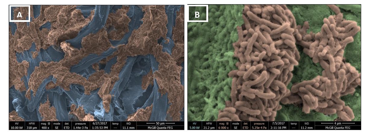
Above. Colorized SEM images of the Kaltostat™ wound dressing at different magnifications. Colorization was performed with TopoMaps software based on Mountains®. Bacterial biofilms are made visible by colorization in light brown.
Different types of bacteria were easily identifiable in the SEM images, both in isolated forms and in clusters within biofilm. For example, it was possible to observe cocci-type bacteria establishing biofilm. Bacilli-type biofilm and streptococci were also observed. All these types of bacteria were detected for both dressings, the main difference being that they were highly visible in the Kaltostat™ dressing and were less notable in both quantity and frequency in the Xeroform™ dressing.
The images above, colorized using Mountains®- based TopoMaps software1, attest to the presence and extent of bacterial biofilm in a lower magnification view (A) where bacterial biofilm is colored in brown and wound dressing fibers in blue.
Furthermore, a higher magnification view image (B) shows bacterial clusters (colored in brown) as well as the extracellular polysaccharide network (EPS matrix, colored in green) which partially surrounds the bacteria, forming the biofilm. “Colorizing the images helped us to better highlight the presence of bacterial biofilms on SEM images at low magnification” stated Dr Iliescu, “It also helped to enhance the presence of bacterial aggregates (clusters) and the formation of the EPS matrix on SEM images at high magnification.”
Further possibilities in biomedical research
This study allowed the team to show that the scanning electron microscope is undoubtedly an immensely valuable tool for the analysis of wound dressing samples for which it had not been previously used.
This imaging technique permits researchers to visualize and characterize bacterial biofilms, provided that the preparation technique is appropriate and that results are correctly analyzed.
The team was able to observe that while donor sites covered with the commonly used wound dressing Kaltostat™ often became breeding grounds for microorganisms, the dressing with bacteriostatic properties (Xeroform™) hindered bacterial proliferation and accelerated wound healing. In other words, bacterial biofilm formation was more pronounced in the wound dressing without bacteriostatic properties.
1 www.fei.com/software/topomaps-for-materials-science/
Read more
▶ In-situ characterization of the bacterial biofilm associated with Xeroform™ and Kaltostat™ dressings and evaluation of their effectiveness on thin skin engraftment donor sites in burn patients. Monica Iliescu Nelea, Laurence Paek, Lan Dao, Nathalie Rouchet, Johnny I. Efanov, Coeugniet Édouard, Michel A. Danino. Burns, Volume 45, Issue 5, August 2019, Pages 11221130. doi.org/10.1016/j.burns.2019.02.024
Colorize your SEM images in just a few clicks
Quick & easy SEM image colorization explained in this video:
How to colorize SEM images with Mountains® image analysis software
Instruments & software used
Thermo Scientific™ (former FEI) Quanta™ 450 FEG Environmental Scanning Electron Microscope (ESEM™) + TopoMaps software